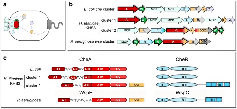
Fig. 2.

Chemosensory clusters identified in the genomic sequence of H. titanicae KHS3. a Schematic representation of the localization and interactions of E.coli chemotaxis proteins. Light green, chemoreceptors; red oval, CheA; dark green square, CheW; lilac, CheZ; light blue oval, CheR; light gray, methylesterase domain from CheB; light orange, receiver domains/proteins (CheB and CheY); dark gray square, flagellar motor. b Gene organization of chemosensory clusters in E. coli, H. titanicae KHS3 and P. aeruginosa (cluster wsp). Each gene is represented as an arrow. Light green, chemoreceptors; red, cheA; dark green, cheW; lilac, cheZ; light blue cheR; light gray, cheB; light orange, receiver domains (in CheB, diguanylate cyclase, CheA and CheY); dark gray, diguanylate cyclase domain. MCPs in H. titanicae KHS3 cluster 1 are RO22_21455, RO22_21470 and RO22_21475. The only MCP in cluster 2 is RO22_21155. c Domain organization of CheA and CheR proteins; A I: Hpt or histidine phosphotransfer domain (P1), A II: CheY/B binding domain (P2), A III: signal transducing histidine kinase, homodimerization domain (P3), A IV: HATPase histidine kinases like ATPase domain (P4), A V: CheW-like domain (P5), A VI, response regulator. R I, methyltransferase domain; R II, S-adenosyl methionine binding; R III, tetratricopeptide repeats
