Fig. 5.

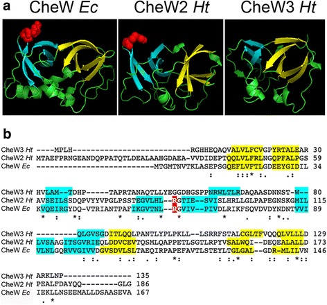
Predicted structure of CheW2 and CheW3 from H. titanicae KHS3. a Comparison between the template and models. Structure of the template CheW from E. coli (PDB accession code 2HO9, left) and the models obtained with SWISSMODEL for CheW2 Ht (middle) and CheW3 Ht (right). The β-strands that form the two β-barrels are colored yellow (subdomain 1) or cyan (subdomain 2). The conserved residue R62 in subdomain 2 of CheW Ec is shown as red spheres. b Clustal Omega alignment of CheW Ec, CheW2 Ht and CheW3 Ht. β-strands are color-coded as in A. Notice that subdomain 2 is incomplete in CheW3 Ht.
