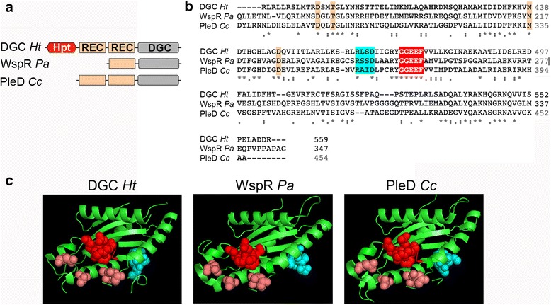
Fig. 6.

Predicted structure of the diguanylate cyclase (chemosensory cluster 2) from H. titanicae. a Schematic representation of cluster 2 diguanylate cyclase from H. titanicae (DGC Ht), WspR from P. aeruginosa (WspR Pa) and PleD from C. crescentus (PleD Cc). A red hexagonal shape indicates the histidine phosphotransfer domain (Hpt; receiver domains (REC) are indicated by light orange rectangles, and diguanylate cyclase catalytic domains (DGC) by gray rectangles. b Clustal Omega alignment of catalytic DGC domains from DGC Ht, WspR Pa and PleD Cc. Active site residues GGEEF are highlighted red, I-site residues RxxD are highlighted cyan, and other conserved residues that have been shown to be involved in activity are highlighted orange (c) Modeled structure for the catalytic domain from DGC Ht compared with the corresponding crystal structures from WspR Pa (PDB accession code 3BRE) and PleD Cc (PDB accession code). Residues shown as spheres are color-coded as in B
