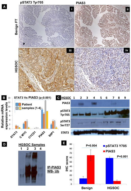
Fig. 1. Characterization of pSTAT3 Tyr705 and PIAS3 expression in benign fallopian tube tissue (Benign FT) and tissue from Grade IV HGSOC patient.
(A) pSTAT3 Tyr705/PIAS3 IHC scoring: benign fallopian tube tissues are characterized by the absence of pSTAT3 Tyr705 (A–i); HGSOC tissue samples are characterized by overexpression of pSTAT3 Tyr705 (A–iii). In contrast, benign fallopian tube tissues show marked expression of PIAS3 in 90–100% of cells (A–ii) whereas only <30% of HGSOC cells express PIAS3 (A–iv). This inverse relationship in pSTAT3 Tyr705 and PIAS3 holds true throughout our results. (B) In order to analyze the expression of PIAS3 along with pSTAT3 Tyr705 and its associated genes at the level of mRNA, we proceeded with extracting RNA from FFPE ribbons of benign FT tissues (4 different patients), which was converted to cDNA and subjected to real time PCR. Figure 1B displays the relative expression of PIAS3 is 2 to 4 folds higher in the benign FT tissue as compared to STAT3, c-Myc, StIP1 and Cyclin D1, which are present at lower levels (p≤0.005). (C) Protein lysates from 9 HGSOC tissues collected from consented patients was subjected to Western Blot analyses and probed with PIAS3, pSTAT3 Tyr705, pSTAT3 Ser727 and total STAT3. Only 3 patients show low expression of PIAS3 and it is entirely absent from the rest of the samples. There is very high pSTAT3 Tyr705 and total STAT3 expression in all the 9 samples, while pSTAT3 Ser727 is rather low. (D) To analyze if the proteasomal-mediated degradation is involved in lowering the levels of PIAS3 in HGSOC tissues, HGSOC tissue lysates were immunoprecipitated (IP) with PIAS3 antibody and immunoblotted (IB) using Ubiquitin. A clear increase in ubiquitination of PIAS3 was evident. (E) Additional scoring of PIAS3 and pSTAT3 Tyr705 expression was completed using IHC in 4 samples. PIAS3 scores were higher for all the benign FT tissues and low for the HGSOC tissues. On the contrary, pSTAT3 Tyr705 scored 5–15% for the benign FT tissues and >70% for the HGSOC tissues. It is noteworthy that there is an inverse relationship between pSTAT3 Tyr705 and PIAS3, which is shown in both mRNA and protein expression levels in both benign fallopian tube tissues and HGSOC. There was a trend of high PIAS3 levels and low pSTAT3 Tyr705 levels in benign tissues, while the inverse was true for HGSOC tissues.

