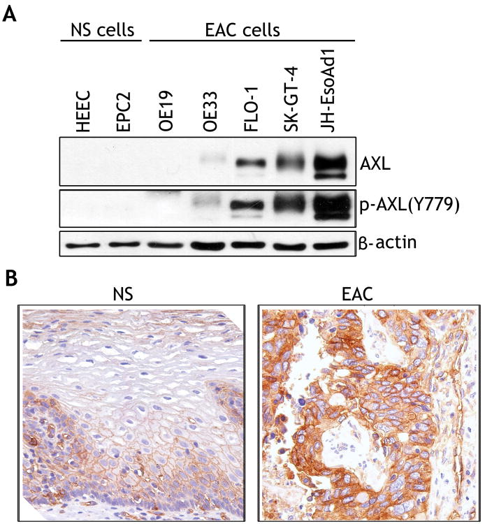
Figure 1. AXL is overexpressed in esophageal adenocarcinomas.
A) Protein extracts from normal esophageal squamous epithelial cell lines (NS) and esophageal adenocarcinoma cell lines (EAC) were subjected to Western blot analysis of AXL and p-AXL(Y779) proteins. B) A representative AXL immunohistochemical staining (IHC) of normal esophageal squamous epithelial tissue sample (NS) (left panel), showing a weak membrane staining (brown color) predominantly in the lower third of the mucosa. A representative AXL IHC staining of a moderately differentiated esophageal adenocarcinoma tissue sample (EAC) (right panel) indicating a strong cytoplasmic and membrane staining.

