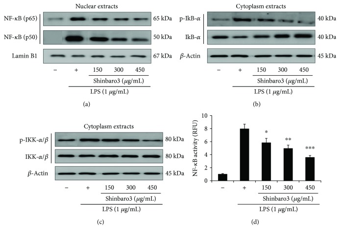
Figure 4.
Effects of Shinbaro3 on NF-κB activation in LPS-stimulated RAW 264.7 cells. RAW 264.7 cells were treated with LPS (1 μg/mL) and Shinbaro3 (150, 300, and 450 μg/mL) for 2 h to investigate protein expression or 6 h for SEAP analysis. Nuclear and cytoplasm extracts were analysed via Western blotting. (a–c) The expression levels of NF-κB (p65 and p50 subunits) (nuclear fraction), p-IκB-α, IκB-α, p-IKK-α, and IKK-α (cytoplasmic fraction) were measured using specific antibodies. Lamin B1 (nuclear fraction) and β-actin (cytoplasmic fraction) were used as an internal control. The data are representative of three separate experiments. (d) Effects of Shinbaro3 on NF-κB transcriptional activity were assessed though a reporter gene assay. Relative fluorescence units (RFU) were measured by using a fluorometer for secreted alkaline phosphatase activity (SEAP). The data are expressed as the mean ± SD (n = 3). ∗p < 0.05, ∗∗p < 0.01, and ∗∗∗p < 0.001 versus LPS treatment alone.

