
Figure 6.
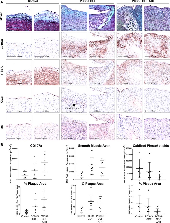
Immunohistochemical analysis of plaques in the right coronary artery. A, Immunohistochemistry staining for CD107a, α‐smooth muscle actin (α‐SMA), CD31, and E06 highlights inflammation, neointimal hyperplasia, endothelial cells, and oxidized phospholipids, respectively, predominantly in gain‐of‐function (GOF) groups. B, CD107a, α‐SMA, and E06 immunostaining areas were quantified on HALO platform (Indica Labs, Corrales, NM), and results were presented as total CD107a‐ or α‐SMA–positive area per plaque area and the percentage of plaque area. Mean and SD are displayed. *P<0.05 compared with control; † P<0.05 compared with proprotein convertase subtilisin‐like/kexin type 9 (PCSK9) GOF receiving standard diet using 1‐way ANOVA, multiple comparison analysis. ATH indicates atherogenic.