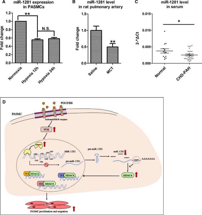
Figure 6.

Reduced miR‐1281 level is identified in hypoxic pulmonary artery smooth muscle cells (PASMCs), rats with pulmonary arterial hypertension (PAH), and patients with PAH. The relative miR‐1281 levels were estimated by real‐time quantitative reverse transcription–polymerase chain reaction in PASMCs exposed to normoxia (21% O2) or hypoxia (3% O2) for 12 and 24 hours (A), pulmonary arteries individually collected from 4 rats with monocrotaline (MCT)–induced PAH and 4 controls (rats received intraperitoneal injections of normal saline; B), and serum individually collected from 13 healthy participants and 29 patients with coronary heart disease (CHD)–PAH (C). D, Proposed regulatory model of phosphatidylinositol 3‐kinase (PI3K)–DNA methyltransferase 1 (DNMT1)–miR‐1281–histone deacetylase 4 (HDAC4) axis. Upward arrow indicates upregulation or activation. Downward arrow indicates downregulation. N.S. indicates not significant; PDGF, platelet‐derived growth factor; and TF, transcription factor. *P<0.05, **P<0.01 compared with normoxia, saline or normal control. Exact P values in consecutive order are: 0.00332, 0.001079, 0.024024.
