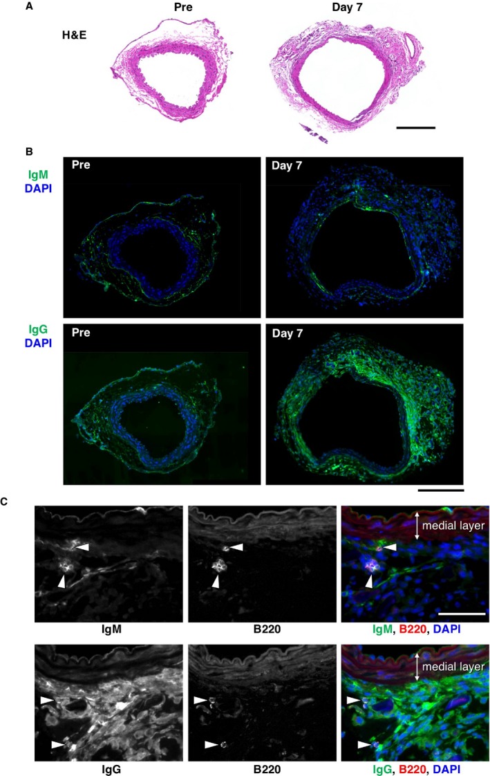
Figure 2.

Deposition of immunoglobulins in mouse AAA. A, Tissue sections are shown for mouse aorta without (Pre) and with (Day 7) CaCl2 challenge by hematoxylin and eosin staining (H&E). Bar 200 μm. B, Deposition of IgM or IgG was detected by immunofluorescence staining (green color) and shown with nuclear staining (DAPI, blue color). Bar 200 μm. C, Deposition of IgM or IgG is visualized by immunofluorescence staining (green color in the rightmost panels) with B‐cell marker B220 staining (arrowheads, red color in the rightmost panels) in aortic tissue 7 days after CaCl2 challenge. Bar 50 μm. AAA indicates abdominal aortic aneurysm; DAPI, 4′,6‐Diamidino‐2‐Phenylindole, Dihydrochloride.
