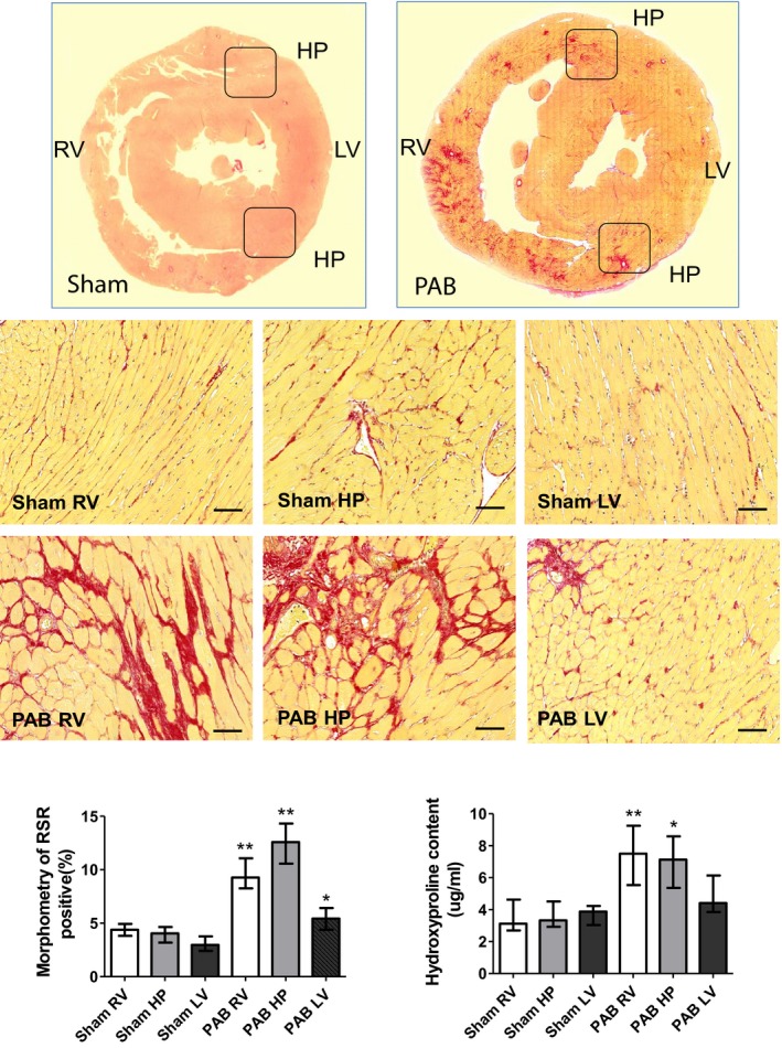
Figure 3.

Representative Picrosirius red (PSR) staining of rat hearts 6 weeks after sham and pulmonary artery banding (PAB) procedures. PSR staining demonstrates that PAB‐induced right ventricular (RV) pressure load is associated with a remarkable accumulation of PSR‐positive collagen and myocyte hypertrophy in the RV and septal hinge‐point (HP) regions. In contrast, the left ventricle (LV) displays only disseminated foci of PSR‐positive material (top panel). Low magnification of heart histological cross‐sections derived from sham and PAB animals and stained with PSR (middle panels). Higher magnification of representative sections derived from the RV, hinge‐point (HP), and LV heart regions (bottom panels). Bars=50 μm. The bar graphs depict morphometric quantification of the areas occupied by PSR‐positive collagen, as well as values of hydroxyproline content. Values are expressed as medians and interquartile range (n=5). *P<0.05, **P<0.005 vs sham.
