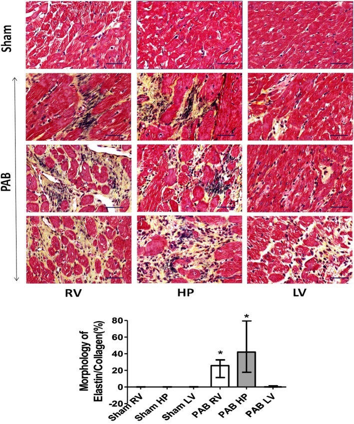
Figure 4.

Representative Movat's staining of rat hearts 6 weeks after sham and pulmonary artery banding (PAB) procedures. Movat's staining depicting collagen I (yellow) and elastin (black) marking differences in regional cardiac fibrosis and extracellular matrix composition in fragments dissected from the right ventricle (RV), hinge‐point (HP), and left ventricle (LV) heart regions of sham and PAB rats (top). There is increased elastin deposition most prominently at the septal HP. Bars=50 μm. The bar graphs depict morphometric quantification of areas occupied by Movat's‐positive elastin and collagen (bottom). Values are expressed as medians and interquartile range (n=5). *P<0.05 vs sham.
