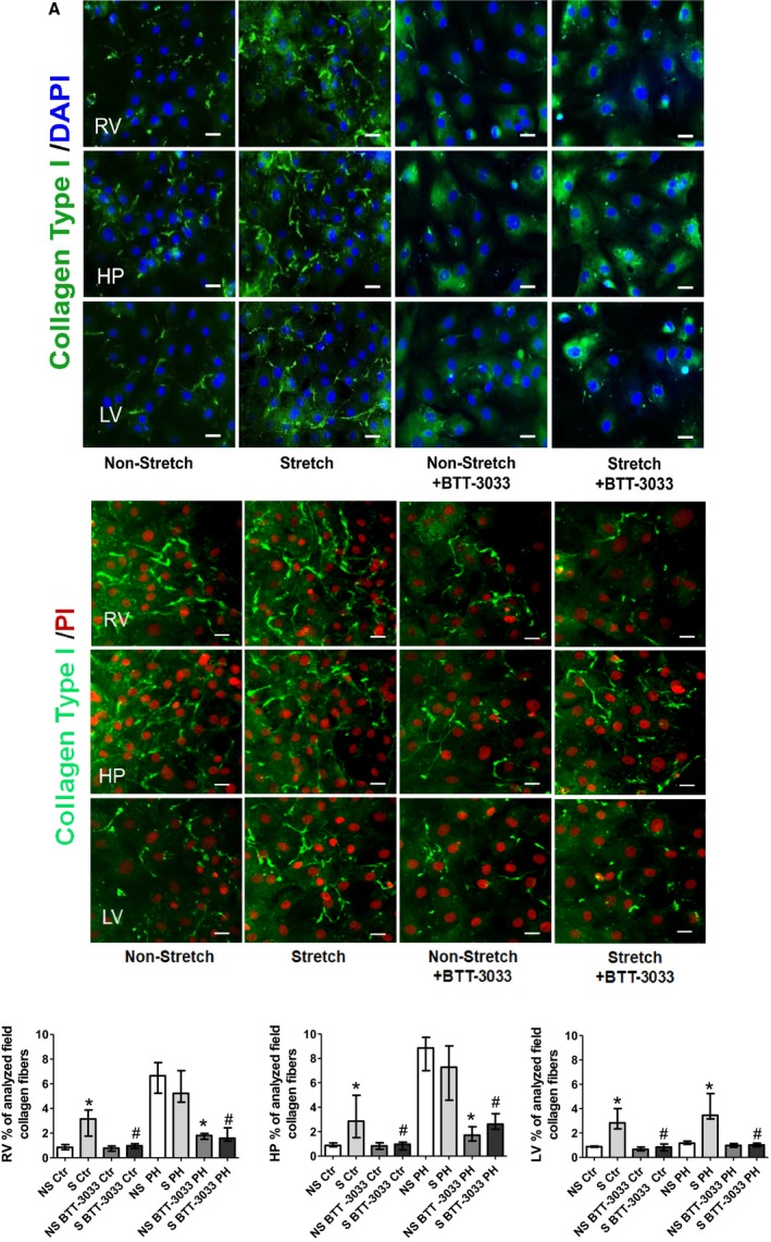
Figure 8.

Cyclic mechanical stretch (24 hours long) of cultured cardiac fibroblasts isolated from the indicated heart regions. A, From control rats, induced a strong upregulation of immune‐detected collagen I deposition, compared with the nonstretched counterparts. This phenomenon has been suppressed in parallel cultures exposed to integrin inhibitor (BTT‐3033) (top panels). B, From pulmonary hypertension rats, indicated strong immune‐detected collagen I deposition in non‐stretched and stretched cells of right ventricular (RV) and hinge‐point (HP) regions, but not in left ventricle (LV). C, Representative fields of cardiac fibroblasts immune stained with collagen type I (green) and nuclear 4′,6‐diamidino‐2‐phenylindole (DAPI) staining (blue)/propidium iodide (PI) (red). Bar=50 μm. The bar graph represents the quantification data of the percentage of analyzed collagen fibers per field. Values are expressed as medians and interquartile range (n=4). Ctr indicates control; NS, nonstretch; PH, pulmonary hypertension; and S, stretch. * P<0.05 vs NS group; # P<0.05 vs S group.
