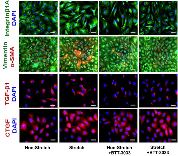
Figure 9.

Representative micrographs depicting cultures of cardiac fibroblasts isolated from right ventricle (RV) that were either kept still or subjected to 24‐hour‐long mechanical stretching in the presence or absence of integrin inhibitor (BTT‐3033). The parallel cultures were immune stained with specific antibodies recognizing integrin‐β1A (green), vimentin (green), α‐smooth muscle actin (α‐SMA) (red), transforming growth factor (TGF)‐β1 (red), and connective tissue growth factor (CTGF) (red), combined with blue 4′,6‐diamidino‐2‐phenylindole (DAPI) nuclear staining. Bars=50 μm.
