
Figure 4.
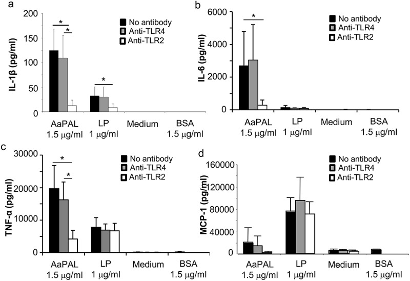
TLR2 dependency of AaPAL-induced cytokine production from the mouse macrophage cell line RAW 264.7. Before incubation with AaPAL for 24 h, the cells were treated with 4 µg of anti-mouse TLR2 (white), anti-mouse (grey), isotype controls (not shown), or no antibody (black) for 2 h. LP denotes synthetic bacterial lipopeptide Pam3CSK4 (EMC Microcollections GmbH). Bovine serum albumin (BSA) was added to the experiments because the AaPAL preparation contained small amounts of BSA as a contaminant [28]. Supernatant was collected, and the produced amounts of the cytokines (a) IL-1β, (b) IL-6, (c) TNF-α, and (d) MCP-1 were determined using ELISA kits (R&D Systems). The graph shows means and SD from three separate experiments. * denotes a statistically significant difference (p < 0.05; Mann-Whitney U-test) between bars connected by the line.