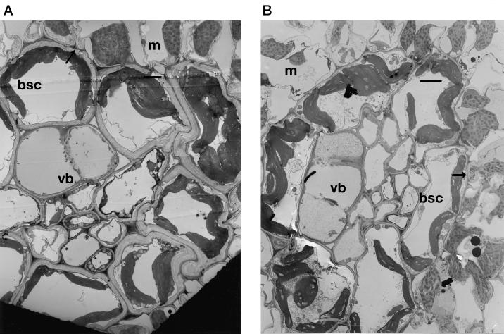
Figure 4.
Transmission electron micrographs of leaf sections showing bundle sheaths from S. bicolor grown at either 350 (a) or 700 μL L−1 CO2 (b). bsc, Bundle sheath cell; m, mesophyll; vb, vascular bundle. Scale bar = 15 μm (both micrographs). Bundle sheath cell walls (indicated by arrows) were approximately twice as thick in ambient relative to elevated CO2 grown plants.

