
Fig. 2.
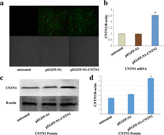
Expression of CNTN1 in Hs578T cells. a Immunocytochemical staining 48 h after transfection, b Real-time RT-PCR: Expression of CNTN1 was significantly increased in CNTN1-transfected cells (P<0.05). c Western blot. CNTN1 was overexpressed compared with controls (P<0.05), d Western blotting quantitation