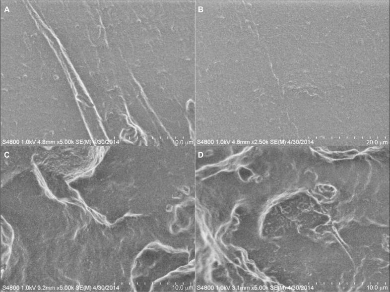
Figure 5.
SEM micrographs of tensile-fractured PDMS and PDMS–HNT surfaces. (A, B) PDMS, (C, D) PDMS–10% HNT. Different fracture patterns were noticed for PDMS and PDMS–10% HNT. Normal PDMS appeared to fracture smoothly, while the HNT-loaded versions displayed rougher fracturing patterns.
Abbreviations: HNT, halloysite nanotube; PDMS, polydimethylsiloxane; SEM, scanning electron microscopy.

