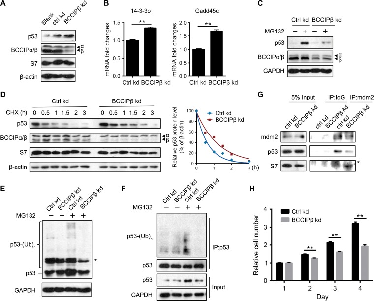
Figure 4.
BCCIPβ depletion triggers ribosomal stress. (A) Total proteins from control and BCCIPβ kd cells were extracted and subjected to western blotting. (B) Total mRNA levels of p53 target genes in control and BCCIPβ kd cells were determined by real-time PCR. (C) After 4 h incubation with DMSO or MG132 (50 μM), control and BCCIPβ kd cells were harvested, and target proteins were detected by western blotting. (D) Control and BCCIPβ kd cells were exposed to the protein synthesis inhibitor CHX (10 mg/ml) for various times, and target proteins were detected by western blotting (left panel). Relative p53 levels at different time points were plotted (right panel). (E and F) Ubiquitinated p53 in control and BCCIPβ kd cells was detected either directly by western blotting with p53 antibody (E) or separated by immunoprecipitation with p53 antibody, followed by detection via western blotting with ubiquitin antibody (F). Asterisk indicates nonspecific band. (G) Cell lysates from control and BCCIPβ kd cells were immunoprecipitated with mouse IgG or MDM2 antibody, followed by western blotting. Asterisk indicates nonspecific band. (H) The proliferation of control and BCCIPβ kd U2OS cells was examined by CCK-8. The results are representative of three different experiments. **P < 0.01.

