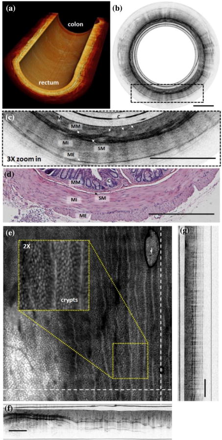
Fig. 3.

(a) Cutaway view of a reconstructed 3D OCT image of a section of mouse colon and rectum along a 2.4 cm catheter pull-back distance with a 20 μm pitch at 20 fps. (b) Representative cross-sectional image of mouse rectum. (c) 3× zoomed-in region of the dotted box in (b), with a representative histology micrograph shown in (d). (e) En face projection image constructed by axial intensity summation over a 4.8 mm × 1.23 mm × 5 mm radial × depth × pull – back) field of view. Crypts and vessel structures are more readily visible. Inset: Enlarged view of the yellow box region. (f)–(g) Cross-sectional image along the dashed lines in (e), which correspond to the radial and longitudinal (pull-back) direction, respectively. M, mucosa; MM, muscularis mucosae; SM, submucosa; MI, muscularis interna; ME, muscularis externa; C, crypts. Scale bar is 500 μm.
