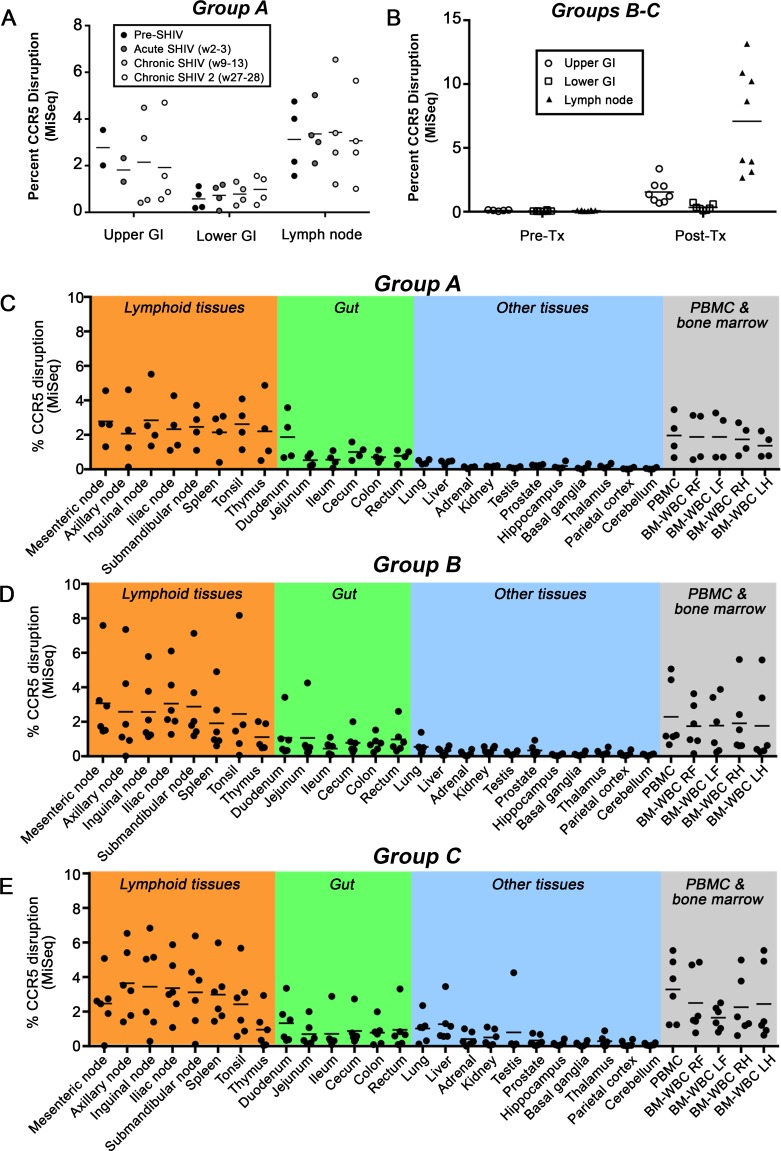
Fig 3. ΔCCR5 cells persist in tissues of SHIV+ animals.
Tissues were collected longitudinally (figure panels [A-B]) or at necropsy (panels [C-E]) from transplanted animals, and the percentage of CCR5-edited alleles was quantified from total tissue homogenate by Illumina MiSeq. (A) Duodenum/Jejunum (Upper GI), colon (Lower GI) and peripheral lymph nodes from Group A animals transplanted prior to SHIV challenge. (B) Same as panel A, from Group B-C animals transplanted following SHIV infection and stable suppression by cART. (C) Necropsy tissues from Group A animals as in panel A. (D) Necropsy tissues from Group B animals in panel B that were necropsied following cART withdrawal. (E) Necropsy tissues from Group C animals in panel B that were necropsied while stably suppressed on cART.

