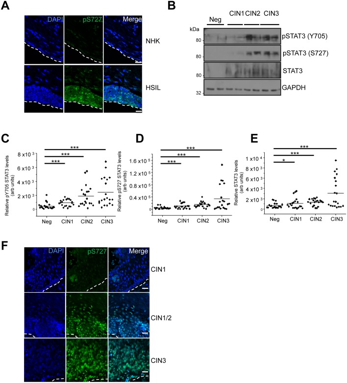
Fig 10. STAT3 expression and phosphorylation is increased in HPV-associated cervical disease.
A) Representative immunofluorescence analysis of sections from organotypic raft cultures of NHK and a W12 cell line presenting with HSIL morphology detecting pS727 STAT3 levels (green). Nuclei were visualized with DAPI (blue) and the white dotted line indicates the basal layer. Images were acquired with identical exposure times. B) Representative western blots from cytology samples of CIN lesions of increasing grade analysed with antibodies specific for phosphorylated (Y705 and S727) and total STAT3 levels. GAPDH served as a loading control. C-E) Scatter dot plot of densitometry analysis of a panel of cytology samples. 20 samples from each clinical grade (neg, CIN I-III) were analysed by western blot and densitometry analysis was performed using ImageJ. Phosphorylated STAT3 levels were first normalised against total STAT3 levels before normalising against protein levels using GAPDH as a loading control. F) Representative immunofluorescence analysis of tissue sections from cervical lesions of increasing CIN grade. Sections were stained for pS727 STAT3 levels (green) and nuclei were visualized with DAPI (blue). Images were acquired with identical exposure times. Scale bar, 20 μm.

