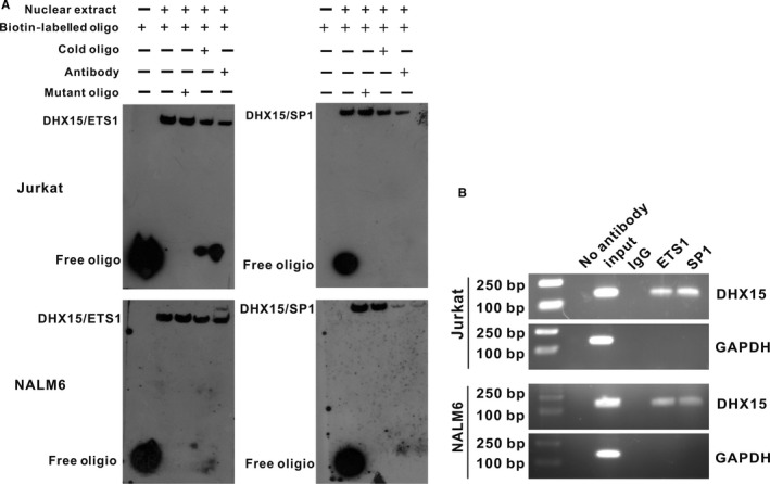
Figure 3.

EMSA and ChIP analyses of ETS1 and SP1 binding to the DHX15 promoter. A, EMSA of ETS1 (left) and SP1 (right). The 5′‐biotin end‐labelled probe was incubated in the absence (lane 0) or presence (lane 1) of nuclear extracts from Jurkat and NALM6 cells. A cold mutated probe (lane 2) and cold probe (lane 3) were used as competitors at concentrations that were in a 100‐fold molar excess to the biotin‐labelled probe. Supershift assays were performed with 4 μg of a specific antibody against ETS1 or SP1 (lane 4). B, Equal amounts of Jurkat and NALM6 chromatin were immunoprecipitated with antibodies for ETS1 and SP1 and subsequently quantified through agarose gel electrophoresis using a primer set specific for the basal region (−181 to −36 bp). Moreover, immunoprecipitated DNA was amplified using a primer set specific to the off‐target region (GAPDH) shown in the lower panel as a negative control
