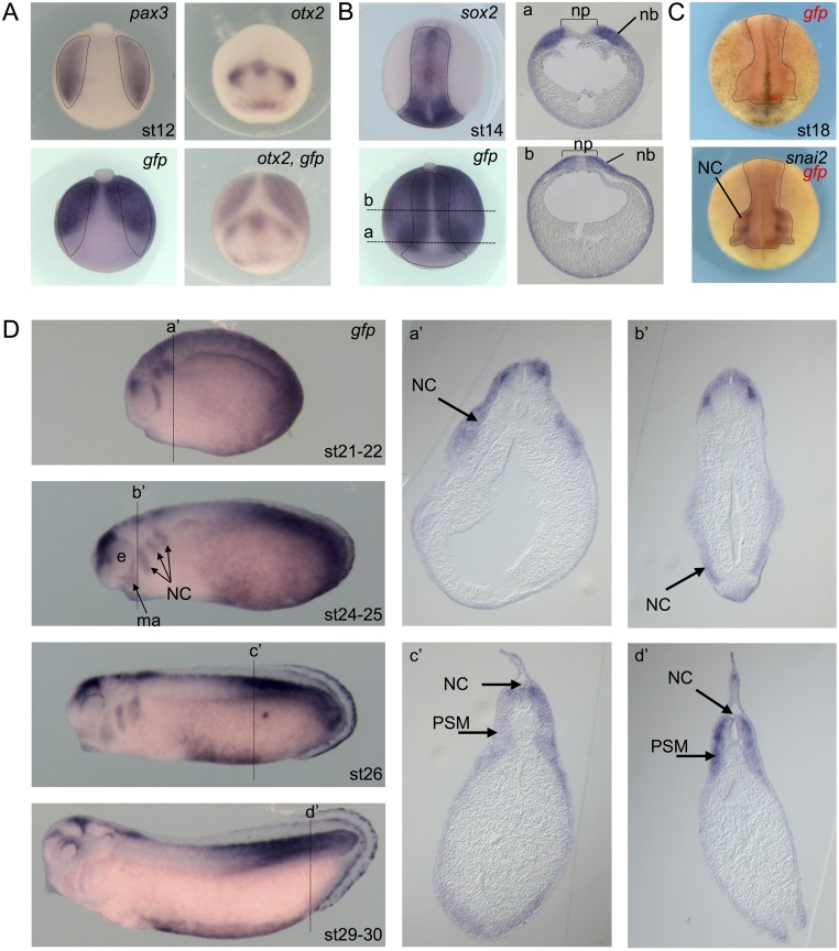
Fig 6. Wnt activity during neural crest formation.
(A) Dorsal view of a stage 12 Tg(pbin7Lef-dGFP) embryo hybridized with pax3 or gfp probes (posterior side is up). Dotted shapes delineate the presumptive neural border on both sides. Anterior view of a stage 12 Tg(pbin7Lef-dGFP) embryo hybridized with otx2 probe alone or otx2 and gfp probes together (dorsal side is up). (B) Dorsal view of a stage 14 Tg(pbin7Lef-dGFP) embryo hybridized with sox2 or gfp or both probes. Dotted shapes delineate the neural plate. The a and b dotted lines indicate the level of shown transverses sections. (C) Anterior views of a stage 18 Tg(pbin7Lef-dGFP) embryo first hybridized with probe against gfp and secondarily with probe against snai2. Dotted lines delineate gfp staining. (D) In situ hybridization against gfp on stage 21–22, 24–25, 26 and 29–30 Tg(pbin7Lef-dGFP) embryos (lateral views). For each stage, the dotted line indicates the level of the shown transverse section (a’-d’). ma: mandibular arch; nb: neural border; NC: migrating neural crest cells; np: neural plate; PSM: posterior presomitic mesoderm.

