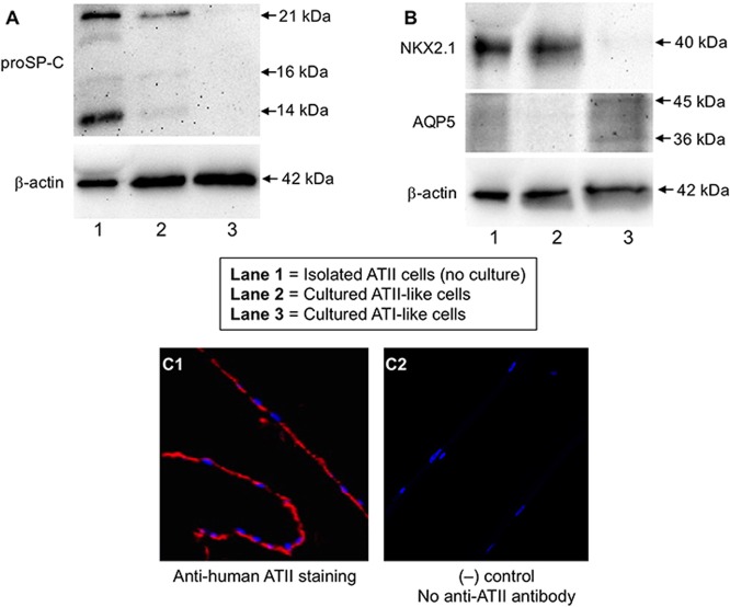
Fig 2. Confirmation of alveolar type II (ATII)-like cells cultured at the air-liquid interface.
Alveolar epithelial cells isolated from a donor were cultured for 7 days to differentiate into ATII-like cells. The cultured ATII-like cells, isolated (non-cultured) ATII cells (positive control) and cultured AT1-like cells from the same donor were processed for Western blot of proSP-C (A), NKX2.1 and AQP5 (B). Additional bands were observed for pro-SP-C (predicted size: 21 kDa) and AQP5 (predicted size: 36 kDa), which is likely due to post-translational modifications, post-translational cleavages, and/or other experimental factors. Cells on the transwell membrane were embedded in paraffin and cut into a 5 μm thickness section for immunofluorescent staining of ATII-like cells (C1) using an anti-ATII cell antibody as described in the Methods section. C2: the negative control—no addition of an anti-ATII cell antibody during immunofluorescent staining. The red and blue colors indicate the cytoplasm and nuclei of ATII-like cells, respectively.

