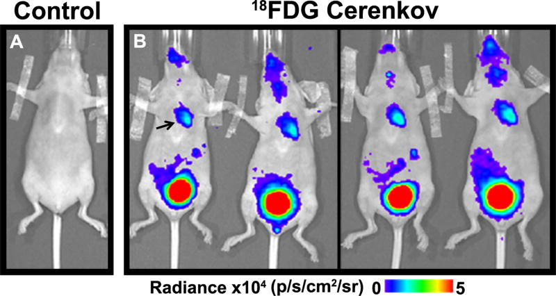
Figure 6. Cerenkov luminescence imaging of the Heart in vivo with 18F-FDG.
(A) No signal was seen in the thorax after saline injection. (B). Robust signal in the thorax (arrow) was seen in the mice injected with FDG, consistent with glucose uptake in the heart. Renal excretion of the probe resulted in an extremely strong signal from the bladder.

