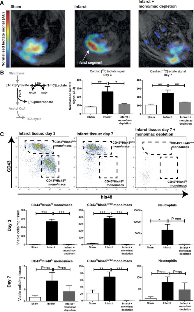
Figure 1.

Hyperpolarized lactate generation in a rodent model of cryoinfarction. A and B, Hyperpolarized MRI demonstrated intense [1-13C]lactate signal at both days 3 and 7 post-experimental myocardial infarction, which was normalized in rats undergoing pharmacological macrophage depletion (n=6 rats assigned per time point per group [n= 36] to give 4–6 evaluable datasets per time point per group; mean±SEM; statistical comparison by 1-way ANOVA with the Holm–Sidak correction for multiple comparisons). Higher lactate signal is detected from the anterior portion of the heart because of some inhomogeneity from the 13C surface coil; lactate signal detected from the chest wall may reflect inflammation in the healing incision. C, Flow cytometric analysis of infarct tissue confirmed predominantly inflammatory monocyte/macrophage phenotype (CD43lohis48hi) at day 3 and predominantly reparative (CD43hihis48lo/int) phenotype at day 7 and that both subsets were depleted after the administration of clodronate liposomes. *P≤0.05, **P≤0.01, ***P≤0.001. LDH indicates lactate dehydrogenase; NADH, nicotinamide adenine dinucleotide; PDH, pyruvate dehydrogenase; and TCA, tricarboxylic acid.
