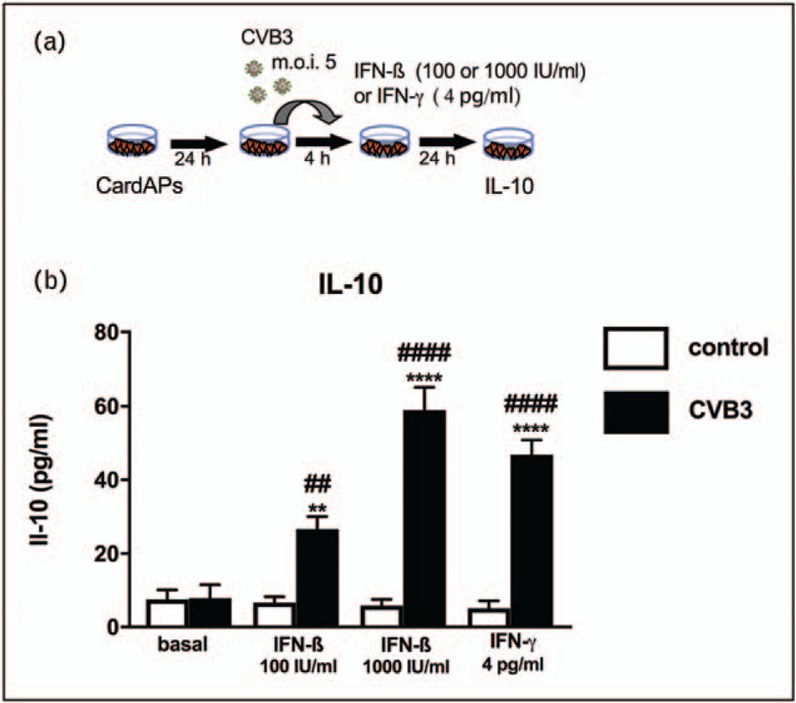
FIGURE 2.

Impact of IFN-ß and IFN-γ on antiviral IL-10 expression of coxsackievirus B3-infected CardAPs. (a) Experimental design illustrating how CardAPs 24 h after plating were infected with coxsackievirus B3 at a multiplication of infection of 5 and 4 h after infection supplemented with/out 100 or 1000 IU/ml of IFN-ß or 4 pg/ml of IFN-γ. Twenty-four hours later, supernatant was collected for subsequent IL-10 analysis via ELISA. (b) Bar graphs represent the mean ± SEM of IL-10 in the supernatant of control CardAPs (open bars) or coxsackievirus B3-infected CardAPs (closed bars) supplemented with/out IFN-ß or IFN-γ, as indicated, with n = 4–6/group and ∗∗P < 0.01 and ∗∗∗∗P < 0.0001 versus respective control group and ##P < 0.01 and ####P < 0.0001 versus the basal coxsackievirus B3 group.
