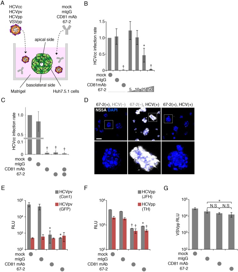
Figure 3. The anti-OCLN mAb 67-2 blocks HCV infection in 3D culture.
(A) Illustration of Huh7.5.1 cell culture system using matrigel-embedded 3D methods to verify HCV infection. (B, C) Huh7.5.1 cells grown in matrigel were exposed to HCVcc and either mock, mIgG (50 μg), anti-CD81 mAb (2.5 μg), the mAb 67-2 (50 μg) or both mAbs, and total RNA from the cells was subjected to real-time RT-PCR analysis for the NS5A expression. The infection levels were expressed relative to the amount of the mock-treated cells, which was taken as 1. (D) Huh7.5.1 cells grown in matrigel were treated with mIgG or 67-2 in the presence or absence of HCVcc, and were subjected to immunostaining for NS5A. Bar, 20 μm. (E–G) Huh7.5.1 cells exposed to mock, mIgG, anti-CD81 mAb or 67-2 in matrigel were subjected to infection assay for HCVpv (E), HCVpp (F) and VSVpp (G). Relative luminescence units (RLUs) were calculated based on the values of VSVpv-infected cells. All values represent the mean ± SD (error bars; n = 5 except for C [n = 6]). Statistic values were determined compared with those in the mIgG-treated cells; *P < 0.01, †P < 0.001. N.S, not significant.

