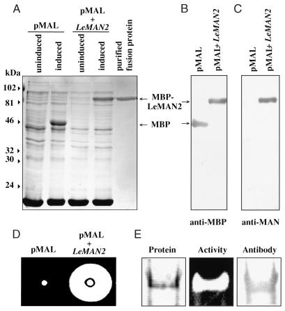
Figure 2.
Overexpression of the protein encoded by LeMAN2 cDNA. A, Protein profiles visualized by Coomassie Blue staining of the lysates of induced or uninduced bacterial cells containing the empty vector (pMAL) or the vector containing LeMAN2 cDNA insert (pMAL + LeMAN2) and the purified fusion protein. The positions of maltose-binding protein (MBP) and the recombinant fusion protein (MBP-LeMAN2) are indicated by the arrows on the right. Molecular masses (kD) are shown on the left. B and C, Immunoblots of the lysates of induced bacterial cells without (pMAL) or with (pMAL + LeMAN2) the cDNA insert, probed with anti-maltose-binding protein antibody (anti-MBP) and anti-M3 mannanase antibody (anti-MAN; Nonogaki et al., 1995), respectively. D, Gel diffusion assays for mannanase activity of the lysates from induced bacterial cells without (pMAL) or with (pMAL + LeMAN2) the cDNA insert. The activity is logarithmically proportional to the size of the cleared area on the gel plate (see Fig. 4). E, Native PAGE of purified maltose-binding protein-LeMAN2 fusion protein. Left, Coomassie-stained protein; center, gel assay for endo-β-mannanase activity; right, immunoblot probed with anti-M3 mannanase antibody.

