
Figure 5.
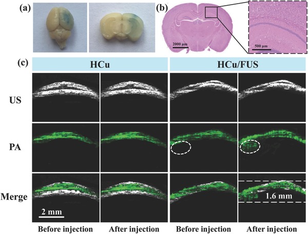
a) EB dye staining of the mouse brain after FUS‐induced BBB opening (Left: an aerial view of a whole brain; Right: brain tissue section). b) Left: The corresponding H&E staining of brain tissue section; Right: Representative microphotograph at high magnification of the boxed area in the left. c) Ultrasound (US), PA, and their overlay images of orthotopic brain tumors acquired before and after intravenous injection of HCu nanosystems without or with FUS‐induced BBB opening.