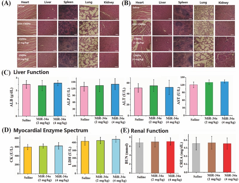
Figure 8.

A) HepG‐2 and B) MDA‐MB‐231 tumor bearing mice intravenously injected with saline, GSH‐CMINs, CMINs (2 mg kg−1 of miR‐34a), and CMINs (4 mg kg−1 of miR‐34a) were sacrificed at 22 d post‐treatment. Representative hematoxylin and eosin (H&E) stained histological images of tissue sections from major organs (heart, liver, spleen, lung, and kidney). Scale bars in all images represent 100 µm. Serum biochemical examination of CMINs‐treated mice. C) Liver function, D) myocardial enzyme spectrum, and E) renal function. ALB, albumin; ALP, alkaline phosphatase; ALT, alanine aminotransferase; AST, aspartate transaminase; CK, creatine kinase; LDH, lactate dehydrogenase; BUN, blood urea nitrogen; CREA, creatinine.
