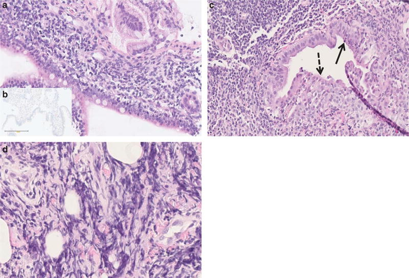
Figure 2.
Pancreas histopathology in pediatric autoimmune pancreatitis (AIP). Representative histopathologic features on pancreas core biopsies in children with autoimmune pancreatitis (hematoxylin and eosin (H&E) staining). (a) Dense lymphoplasmacytic infiltration of the major papilla (original magnification ×20). (b) Negative IgG4 staining of the plasma cells (original magnification ×20). (c) Granulocytic epithelial lesion (arrow) and focal epithelial duct destruction (dashed arrow). The chorion is infiltrated by lymphoplasmatic cells (original magnification ×20). (d) Storiform fibrosis (original magnification ×40).

