
Figure 2.
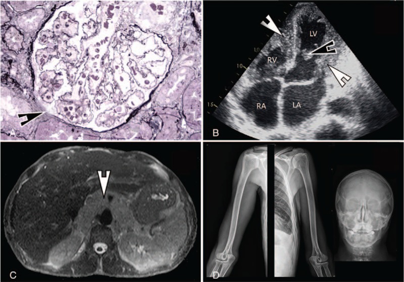
(A) Glomerulus showing wrinkling of capillary walls, parietal arteriolar edema, focal glomerular ischemia, and widening of the subendothelial space (arrows) (Jones’ basement membrane stain, ×400). (B) Trans-thoracic echocardiography showing a nondilated left ventricle with infiltration of both left and right ventricles endocardium (thick arrows) and a left ventricular thrombus (thin arrow). (C) MRI (T2 weighted images) showing a diffuse infiltration of the retroperitoneal space surrounding the aorta and the caval vein with no mass effect. (D) Representative x-ray images (from left to right: right and left humerus and shoulder girdle, skull) showing normal bone structure without pathologic features. LA = left atrium, LV = left ventricle, RA = right atrium, RV = right ventricle.