Figure 5.
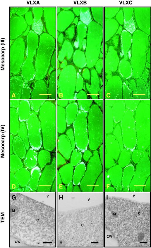
Light and electron microscopy immunolocalization of VLXA, VLXB, and VLXC in 3-week-old soybean pod walls. Note the accumulation of these isoforms in a single cell layer, which confirms the observations seen in Figure 4. Sections were developed using affinity-purified antipeptide antisera for VLXA, VLXB, and VLXC. VLXA, VLXB, and VLXC colocalized to a distinct cell layer in the middle region of the mesocarp. Sections taken from regions III and IV (refer to Fig. 3B) illustrate this cellular localization. A through C, Sections taken from mesocarp region III stained with antisera against VLXA, VLXB, and VLXC, respectively. D through F, Sections taken from mesocarp region IV stained with antisera against VLXA, VLXB, and VLXC, respectively. Scale bar = 50 μm (A–F). G through I, TEM showing subcellular localization of VLXA, VLXB, and VLXC (respectively) in the cytosol of these cells. Scale bar = 500 nm (G–I). C, Cytoplasm; CW, cell walls; M, mitochondria; V, vacuole.

