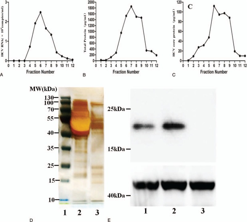
Figure 1.

Purification and identification of infectious HCV particles. (A) Concentrated supernatants isolated from infected cells were subjected to ultracentrifugation using a sucrose step gradient. The collected particles were purified in a continuous 10% to 60% sucrose gradient. HCV RNA levels were determined by quantitative polymerase chain reaction, and the results are expressed as the number of HCV RNA copies per mL. (B) Total protein was determined by BCA, and the results are expressed in μg/mL. (C) HCV core protein levels were assessed by ELISA, and the results were expressed in μg/mL. (D) Representative plot of the gradient fractions showing sodium dodecyl sulfate polyacrylamide gel electrophoresis and silver staining (1: Marker, 2: HCVcc, 3: Purify HCVcc). Sucrose gradient purification removed most of the proteins from the solution of virus particles. (E) Western blot analysis of the HCV core protein with a molecular weight of 20 kDa (1: HCVcc, 2: Purify HCVcc, 3: Control). The HCVcc and purify HCVcc were detected with anti-HCV core protein by western blot, which revealed positive band; control: no band was detected in control. (F) The HCV virions were determined by negative staining and observed by transmission electron microscopy. Representative electron micrographs showing specific purified, negatively stained HCV particles. Scale bar, 200 nm. HCV = hepatitis C virus, HCVcc = HCV cell culture.
