
Fig. 3.
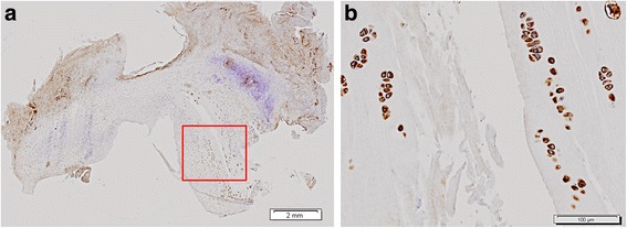
Cell clusters, COMP immunohistochemistry. a Microphotograph of a COMP stained medial meniscus 12 weeks after ACL-transection, where a tear can be seen. b 100×. Cell clusters, showing chondrocyte-like cells, strongly stain for COMP around the meniscal tear from the boxed area in (a)