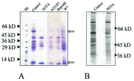
Figure 8.
Soluble proteins and in vivo protein synthesis under different culture treatments: A, 10% (w/v) SDS-polyacrylamide gel showing Coomassie-stained proteins. Arrows indicate prominent protein bands absent under A23187 treatment and Ca2+-chelated/deprived conditions as compared with the control; B, l-[35S]Met labeling of proteins in embryogenic cultures grown under optimal (control) and under Ca2+-chelated (EGTA) differentiation conditions.

