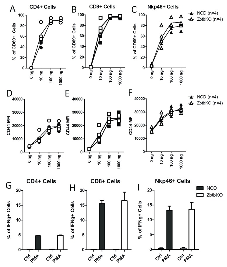
Figure 4. Activation markers and cytokine production in Zbtb32 -/- splenocytes after ex vivo stimulation.
Freshly isolated splenocytes from female NOD.Zbtb32 -/- mice and control littermates were stimulated with the indicated dosages of anti-CD3 for 18 h. Surface expression of CD69 ( A– C) and CD44 ( D– F) was measured on CD4 +, CD8 +, and NKp46 + splenocytes. For IFNγ staining, splenocytes from female NOD.Zbtb32 -/- mice and their control littermates were stimulated with PMA and Ionomycin for 4 hours ( G– I). The percentage of cells positive for IFNγ in CD4 + ( G), CD8 + ( H), and NKp46 + cells ( I) is shown. The data were combined from two independent repeats of this experiment.

