. 2017 Nov 28;9(7):1753–1759. doi: 10.1039/c7sc04243d

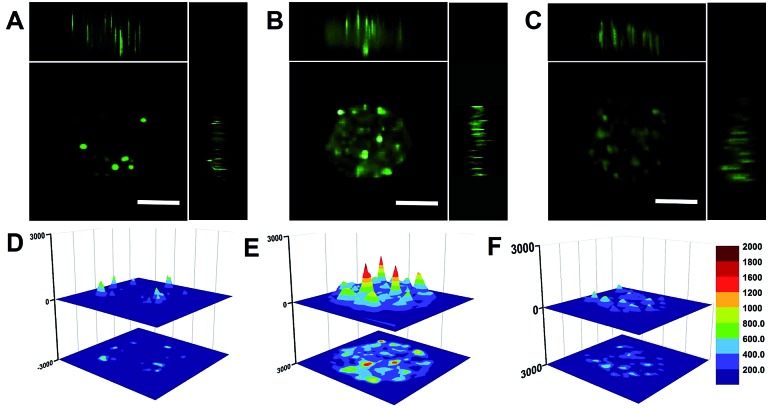
Fig. 5. Penetration of the THP-AuNR nanoprobe in MCF-7 MCTS. (A) Confocal microscopy images of MCTS after incubation with the THP-AuNR nanoprobe for 8 h and without irradiation by an 808 nm NIR laser (x, y, and z directions). (B) Confocal microscopy images of MCTS incubated with the THP-AuNR nanoprobe and treated with an NIR laser (808 nm, 3 min and 0.64 W cm–2) after 24 h (x, y, and z directions). (C) Confocal microscopy images of MCTS incubation with the THP-AuNR nanoprobe and without irradiation by an 808 nm NIR laser after 24 h (x, y, and z directions). (D) Surface plot images of MCF-7 MCTS in A. (E) Surface plot images of MCF-7 MCTS in B. (F) Surface plot images of MCF-7 MCTS in C. Scale bars: 100 μm.

Fig. 5