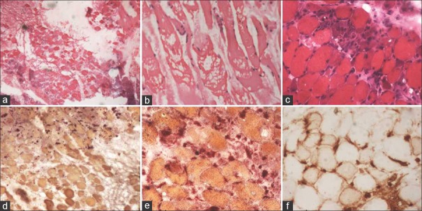
Figure 1.
(a and b) Presence of necrotic muscle fibers on muscle biopsy (H and E, ×40). (c) Many regenerating fibers around the necrotic fibers (H and E, ×100). (d and e) Macrophages around the necrotic muscle fibers take up red color (acid phosphatase ×100). (f) Major histocompatibility complex Class I antigen expression in the muscle fibers

