Figure 1.
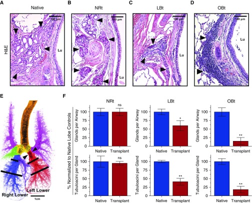
Loss of submucosal glands (SMGs) in ferret pulmonary allografts demonstrating progressive obliterative bronchiolitis (OB) pathology. (A–D) Representative hematoxylin and eosin (H&E)-stained sections of large airways of native lungs (A), transplant lobes with no allograft airway rejection (NRt) (B), transplant lobes with lymphocytic bronchiolitis (LBt) (C), and transplant lobes with OB (OBt) (D). Arrowheads indicate SMGs, and Lu denotes airway lumen. (E) Illustration of normal ferret lung block with color-coded lobar anatomy indicating the trachea (orange), mainstem bronchi (yellow), bilateral upper lobes (pink), right middle lobe (green), right mediastinal lobe (purple), right lower lobe (blue, native control lobe), and left lower lobe (red, experimental transplant lobe). Black bars indicate where sections were obtained for analysis of SMG abundance. Arrowheads indicated where the transplant anastomosis is performed. (F) Quantification of SMGs in ferret allografts (red) versus native control lobes (blue), with respect to number of well-defined glands per airway (top) and number of tubuloacinar structures per gland (bottom) across the same spectrum of disease found in A–D (NRt, n = 3; LBt, n = 6; OBt, n = 5). Data are presented as mean ± SEM. *P < 0.05; **P < 0.01 by Mann-Whitney U test. ns = not significant.

