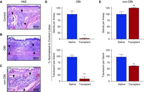
Figure 2.
Loss of submucosal glands (SMGs) in human pulmonary allografts demonstrating progressive obliterative bronchiolitis (OB) pathology. (A–C) Representative hematoxylin and eosin (H&E)-stained sections of large airways from control pulmonary tissue (A), pulmonary allografts with OB (OBt) (B), and pulmonary allografts without OB (non-OBt) from patients who received a lung transplant but died from nonpulmonary complications (C). Arrowheads indicate SMGs, and Lu denotes airway lumen. (D and E) Quantification of SMGs in human pulmonary allografts (red) versus control lobes (blue), with respect to the number of gland clusters per airway (top), and the number of tubuloacinar structures per gland (bottom) in seven OBt allografts (D) and in two non-OBt allografts (E). Blue columns indicate size-matched, age-matched, and sex-matched nontransplanted lung tissue from patients who died of nonpulmonary pathology (n = 7). Data are presented as mean ± SEM. **P = 0.0012 by Mann-Whitney U test. ns = not significant.

