Figure 3.
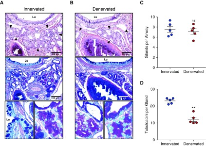
Effects of airway vagal nerve interruption (denervation) on submucosal glands (SMGs) in ferret pulmonary lung lobes. (A and B) Representative hematoxylin and eosin–stained sections (top), serial sections stained with periodic acid–Schiff/Alcian blue (PAS/ab) (middle), and high-magnification PAS/ab images (bottom) of large airways of innervated (nonsurgical) ferret lobes (A), and denervated ferret lobes (B). Arrowheads indicate SMGs, and Lu denotes airway lumen. (C and D) Quantification of the innervated control lobes (blue) and denervated lobes (red) indicating the number of glands (C) and the number of tubuloacinar structures per gland (D). Data are shown as the raw number of glands and tubuloacinar structures in each animal and presented as the mean ± SEM of five animals. No denervated lobes demonstrated histopathological evidence of overt obliterative bronchiolitis. **P = 0.0011 by paired t test. ns = not significant.

