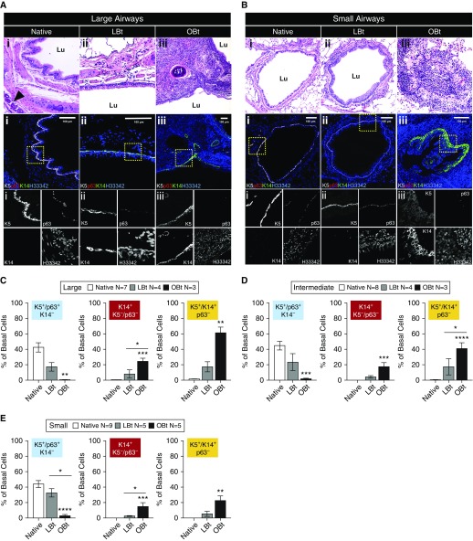
Figure 4.
Changes in the expression of basal cell markers in airways of native and allograft in ferrets with varying allograft disease severity. (A and B) Hematoxylin and eosin–stained sections and serial section immunofluorescence images of (A) large and (B) small airways from (Ai and Bi) native, (Aii and Bii) lymphocytic bronchiolitis allografts, and (Aiii and Biii) obliterative bronchiolitis (OB) allografts. Columns indicate representative airways used for basal cell quantification in the left lower lobe pulmonary allograft in lymphocytic bronchiolitis without OB (LBt) and in OB (OBt) versus mirror-image sections from the native right lower lobe used for quantification of native airway controls. Arrowhead indicates submucosal gland, and Lu denotes airway lumen. (C–E) Quantification of basal cell phenotypic changes in large (C), intermediate (D), and distal (E) airways in ferret allografts as compared with native lobes of the same animals. Sections were stained for K5, p63, and K14 as marked. Statistical analysis was performed by two-way analysis of variance and Tukey multiple comparison post hoc test: *P < 0.05; **P < 0.01; ***P < 0.001; ****P < 0.0001. Data shown are mean ± SEM for number of animals/lobes for each dataset as indicated in the figure.

