
Fig. 1.
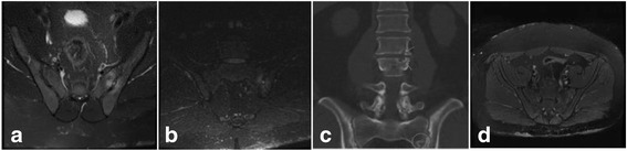
a MRI SIJs (axial) pre-treatment T2 FS demonstrating T2 hyperintense synovitis and marrow oedema b MRI SIJs (coronal) pre-treatment STIR demonstrating acute left sacroiliitis c Conventional CT lumbar spine and SIJs (coronal) with arrows on the L2/3 endplate erosions and circle at the LEFT SIJ erosions d MRI pelvis (axial) post-treatment T2 FS demonstrating resolution of T2 hyperintense synovitis and marrow oedema