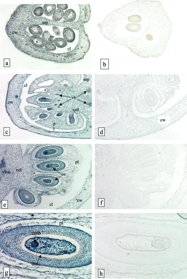
Figure 7.
In situ hybridization of LeFPS to young tomato fruit sections. Bright field micrographs of 7-μm tissue sections from 3.5-mm diameter fruits (a and b), 6-mm (c and d), and 8-mm (e and f) large fruits are shown. g and h, Higher magnification of e and f showing the concentration of labeling in the developing seeds. Sections were hybridized either with a sense (a, c, e, and f) or an antisense (b, d, f, and h) LeFPS1 DIG-labeled RNA probe. The hybridization signal appears as a dark-blue staining and is localized in cells from all fruit tissues. Cw, Carpel wall; sep, septum; ov, ovules; pl, placenta; col, columella; vb, vascular bundles; emb, embryo; end, endosperm; in, integument; din, disintegrating portion of integument. a and b, ×60; c, ×30; d, ×70; e and f, ×20; g and h, ×80.

