
Figure 3.
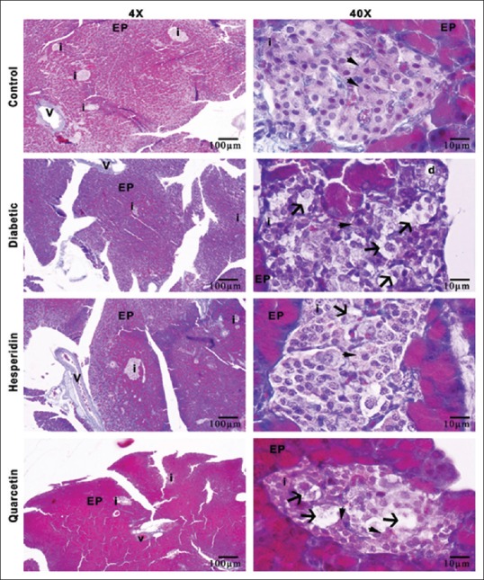
Illustration of histologic structure of rat pancreas tissues for all groups, i: Langerhans islets, EP: Exocrine pancreas, v: Vessel of pancreas, d: Ductus of pancreas, arrowhead; beta cells arrow; degenerated beta cells, Crossman-modified triple staining