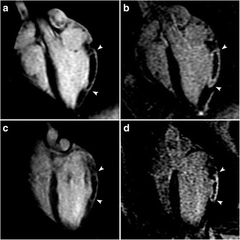
Fig. 3.

Magnetic resonance imaging (MRI) appearance of myocardial infarct (MI) with a significant no-flow region (NF) at post-contrast time points that are used clinically. Representative long-axis early gadolinium enhancement (EGE) (2 min post-contrast) (a, c) and late gadolinium enhancement (LGE) (15 min post-contrast) (b, d) images obtained from animals with reperfused MI (a, b) (top row) and non-reperfused (c, d) (bottom row). MIs are bracketed by arrowheads. The NF can be clearly visualised as a central unenhanced area within the enhanced rim of the MI, even in the late phase in both models
