Fig. 2.

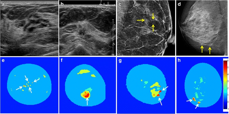
Imaging overview of the four patients with suspicious findings. Top row shows HUS (a, b) and mammography (c, d); bottom row shows the diagnostic index (DI) maps of the MUT exams (e–h) with the colour bar depicting the DI value. a A hypoechoic lesion in the right breast, which is lobulated and ill defined. MUT coded this lesion clearly as benign in orange, corresponding to a DI value of 4 (e, white arrows). Histopathology showed a common ductal hyperplasia. b HUS of the right breast with an irregular lesion and surrounding distortion of the tissue. MUT coded this lesion as malignant in red corresponding to a DI value of > 5 (f, white arrows). Histopathology demonstrated an invasive ductal carcinoma. Mammography shows in (c) regional microcalcifications at 3 o’clock in the left breast (yellow arrows). MUT depicted corresponding confined lesions coded red (g, white arrows). Histopathology confirmed an invasive ductal carcinoma with surrounding DCIS. Mammography shows in panel (d) two superficial suspicious findings (yellow arrows). MUT indicated two findings coded red (h, white arrows). Histopathology confirmed invasive ductal carcinoma (see also Fig. 3)
