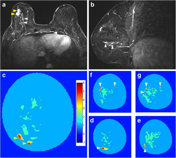
Fig. 3.

Depiction of a multifocal/mulicentric cancer (same case in Fig. 2d, h). Contrast-enhanced T1-weighted fat-saturated MRI in the axial (a) and sagittal reconstructions (b). Depiction of multifocal cancer with two masses in the lower outer quadrant (yellow arrowheads) and additional ductal enhancement in other quadrants (white arrowhead). MUT clearly demonstrates the two masses and coded them correctly in red as indicated in c (yellow arrowheads), but shows also additional small areas with high DI (>5) in the following coronal slices (d–g, white arrowheads). The ductal enhancement was further investigated using an MRI-guided biopsy and the histopathology showed additional DCIS
